Capture, store, retrieve - imaging made easy.
|
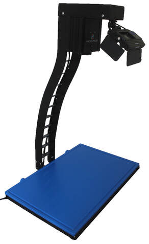
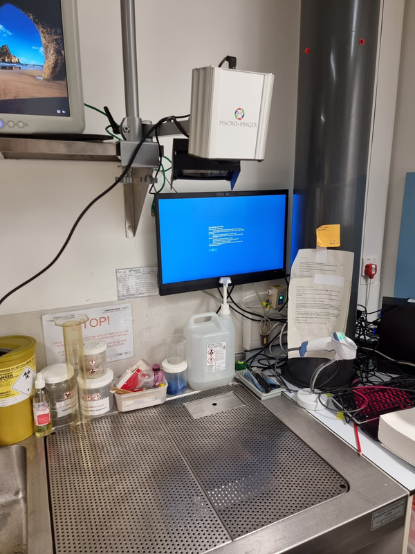
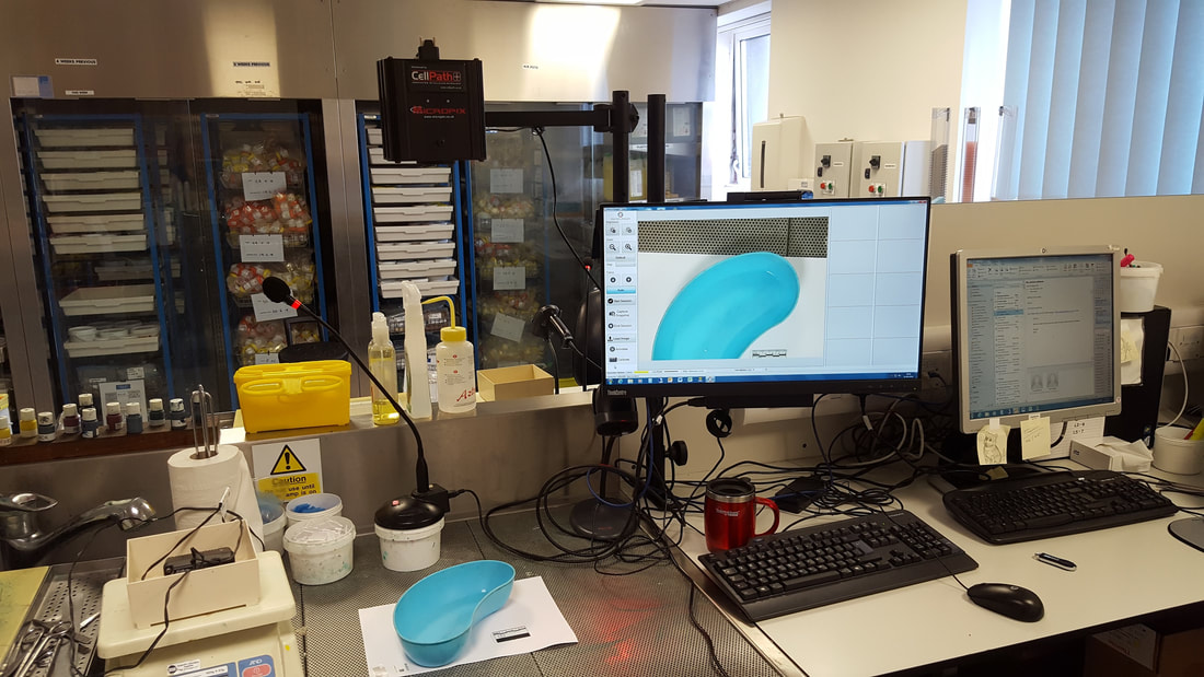
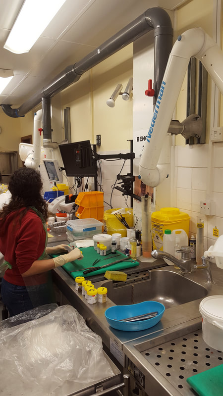
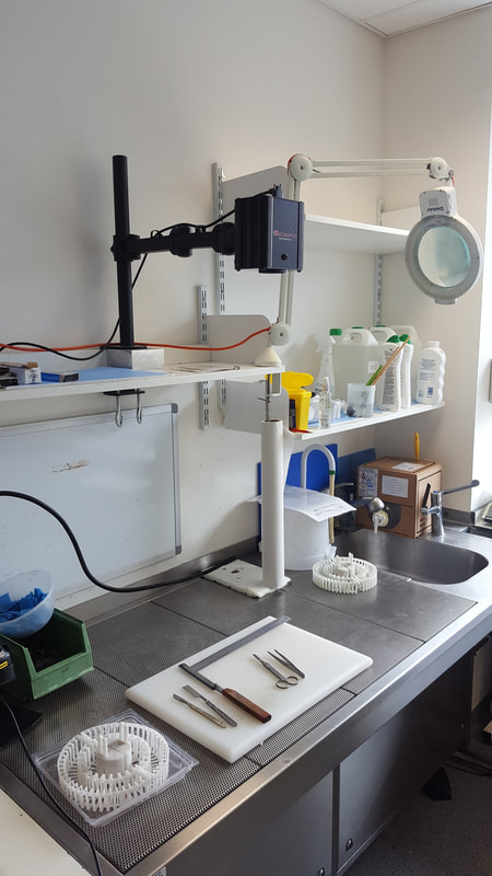
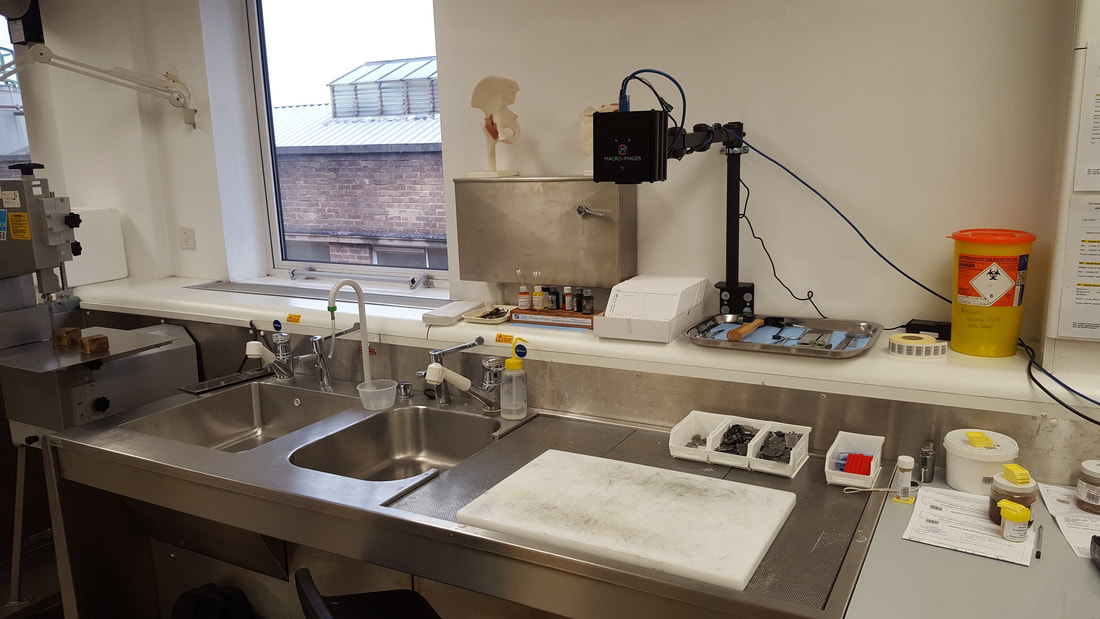
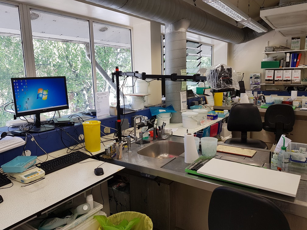
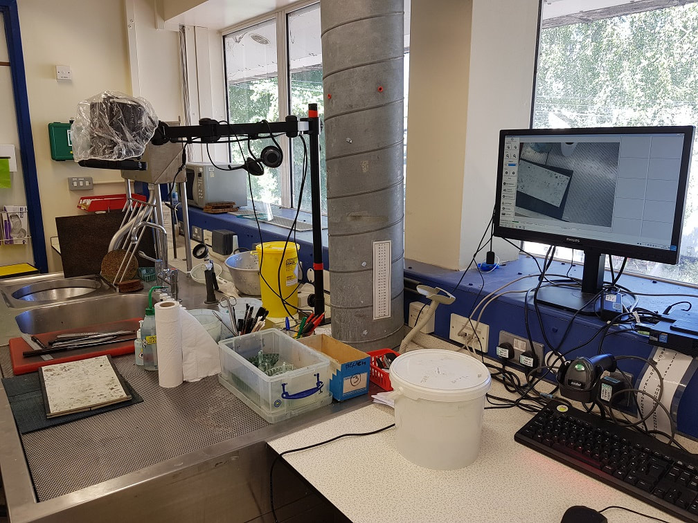
Our Macro-imager system has been designed specifically for use within the cut-up lab of hospital pathology departments. Combining a high-quality, full HD camera with 30x optical zoom with easy-to-use and intuitive software, the Macro-imager makes image capture simple and reliable.
|
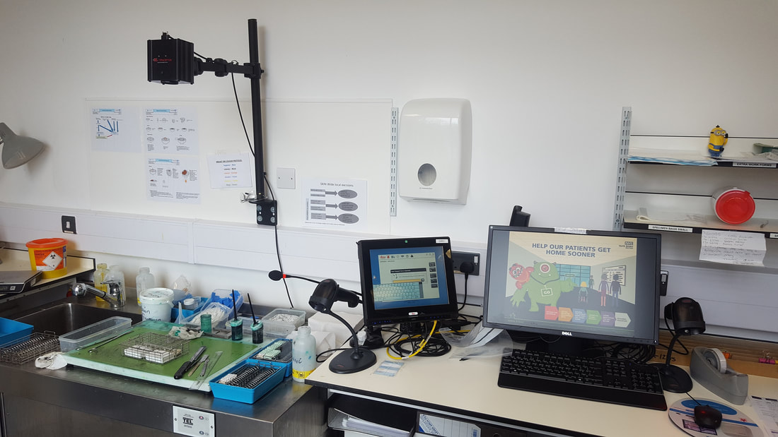
We love our Macro-imager! It is so easy to use with intuitive software and easy on-screen commands, so even the most technophobic of us finds it a doddle. Because it is mounted between cut up benches it is much safer than using a hand-held camera, which is what we had before. The picture quality is fantastic, with an excellent zoom range so you can photograph everything from the largest intact specimens to the smallest areas within them. The annotation facility has gone down a storm – the pathologists love knowing which areas have been sampled specifically, which enables them to correlate the macroscopic findings with the histological features. We wonder now how we coped for so long without it.
Ellen Hayden - Advanced Practitioner in Dissection - Whiston Hospital, Part of St Helens and Knowsley Teaching Hospitals NHS Trust
We have a Macro-imager camera set up overhead at our dissection bench providing the ultimate in convenience for macro photographs. The software is installed on the existing Trust PC on the bench and it takes less than a minute to take your picture. The annotation tool has been a welcome addition so that you can mark your block selection or highlight a specific area straight onto the image for an instant high quality, professional finish without having to edit later. Having the camera overhead also means you don’t have to transport formalin fixed tissue away from the down-flow bench.
Maria Haynes - Dissection Room Lead - Maidstone Hospital


